Knee Extension
Electrode Placement
Application Instruction by Dr. Lucinda Baker
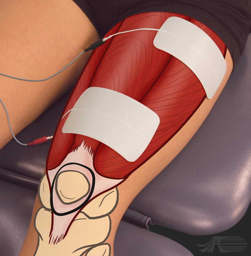
Electrode placement for knee extension. The patella is marked with a circle, distally. The large electrodes are placed along the thigh. The proximal electrode is very proximal and towards the lateral side. The distal electrode is placed either at midline, as shown here, or slightly to the medial side. Too much medial a placement of the distal electrode can create a powerful Vastus Medialis cramp. If this occurs, keep the distal electrode midline to slightly lateral.
Knee Extension
Video Instruction
Audio Transcript:
Electrode placement for knee extension. The patella is marked with a circle, distally. The large electrodes are placed along the thigh. The proximal electrode is very proximal and towards the lateral side. The distal electrode is placed either at midline, as shown here, or slightly to the medial side. Too much medial a placement of the distal electrode can create a powerful Vastus Medialis cramp. If this occurs, keep the distal electrode midline to slightly lateral.
Knee Extension
Muscle Anatomy
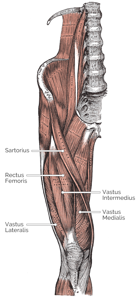
Muscles involved in knee extension:
Rectus Femoris
Origin: Straight head from anterior inferior
iliac spine. Reflected head from groove just
above acetabulum
Insertion: Base of Patella
Vastus Intermedius
Origin: Superior half of anterior and Lateral
surfaces of femur
Insertion: Lateral border of Patella
Vastus Medialis
Origin: Superior half of anterior and medial
surfaces of femur
Insertion: Medial condyle of tibia
Vastus Lateralis
Origin: Superior half of anterior and Lateral
surfaces of femur
Insertion: Lateral border and Base of Patella
Sartorius
Origin: Anterior Superior iliac spine
Insertion: Supero-medial tibia
Other actions: Hip flexion, lateral hip rotation
Knee Extension
Nerve Anatomy
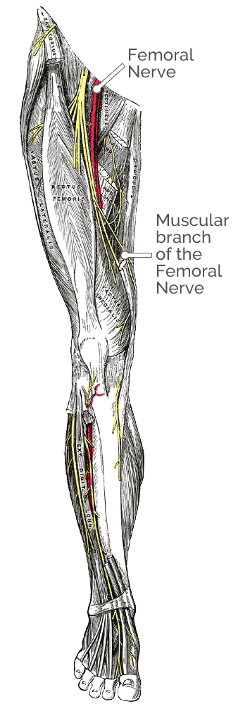
Nerves involved in knee extension:
Rectus Femoris
Nerve innervation: Muscular branches of
femoral nerve
Nerve root: L2, L3, L4
Vastus Intermedius
Nerve innervation: Muscular branches of
femoral nerve
Nerve root: L2, L3, L4
Vastus Medialis
Nerve innervation: Muscular branches of
femoral nerve
Nerve root: L2, L3, L4
Vastus Lateralis
Nerve innervation: Muscular branches of
femoral nerve
Nerve root: L2, L3, L4
Sartorius
Nerve innervation: Femoral nerve
Nerve root: L2, L3, L4
