Knee Extension: Dual Channel Power
Electrode Placement
Application Instruction by Dr. Lucinda Baker
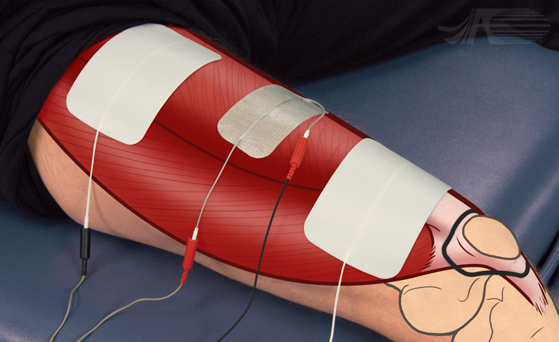
Electrode placement for knee extension dual channel for power activation. Two channels of stimulation are used with an asymmetric biphasic waveform. The negative electrode is placed very proximal and the positive lead is attached to the dual leadwire electrode placed at the mid-thigh. The channel two negative electrode is placed distally, midline to medially. The positive lead is attached to the dual leadwire electrode at the mid-thigh.
Knee Extension: Dual Channel Power
Video Instruction
Audio Transcript:
Electrode placement for knee extension dual channel for power activation. Two channels of stimulation are used with an asymmetric biphasic waveform. The negative electrode is placed very proximal and the positive lead is attached to the dual leadwire electrode placed at the mid-thigh. The channel two negative electrode is placed distally, midline to medial. The positive lead is attached to the dual leadwire electrode at the mid-thigh.
The stimulated contraction is a three out of five, but under ideal circumstances can be increased to three plus or more out of five.
Knee Extension: Dual Channel Power
Muscle Anatomy
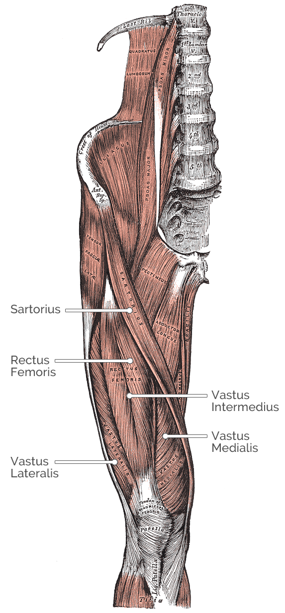
Muscles involved in knee extension:
Rectus Femoris
Origin: Straight head from anterior inferior
iliac spine. Reflected head from groove just
above acetabulum
Insertion: Base of Patella
Vastus Intermedius
Origin: Superior half of anterior and Lateral
surfaces of femur
Insertion: Lateral border of Patella
Vastus Medialis
Origin: Superior half of anterior and medial
surfaces of femur
Insertion: Medial condyle of tibia
Vastus Lateralis
Origin: Superior half of anterior and Lateral
surfaces of femur
Insertion: Lateral border and Base of Patella
Sartorius
Origin: Anterior Superior iliac spine
Insertion: Supero-medial tibia
Other actions: Hip flexion, lateral hip rotation
Knee Extension: Dual Channel Power
Nerve Anatomy
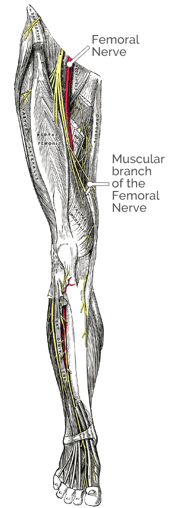
Nerves involved in knee extension:
Rectus femoris
Nerve innervation: Muscular branches of
femoral nerve
Nerve root: L2, L3, L4
Vastus intermedius
Nerve innervation: Muscular branches of
femoral nerve
Nerve root: L2, L3, L4
Vastus medialis
Nerve innervation: Muscular branches of
femoral nerve
Nerve root: L2, L3, L4
Vastus lateralis
Nerve innervation: Muscular branches of
femoral nerve
Nerve root: L2, L3, L4
Sartorius
Nerve innervation: Femoral nerve
Nerve root: L2, L3, L4
