Knee Flexion
Electrode Placement
Application Instruction by Dr. Lucinda Baker
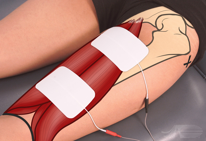
Electrode placement for knee flexion. The greater trochanter and the popliteal fossa are marked. Large electrodes are used to activate this diverse muscle group, with a symmetric biphasic waveform. The proximal electrode is placed medially to activate all of the hamstrings near their origin. The distal electrode is placed about midline.
Knee Flexion
Video Instruction
Audio Transcript:
Electrode placement for knee flexion. The greater trochanter and the popliteal fossa are marked. Large electrodes are used to activate this diverse muscle group, with a symmetric biphasic waveform. The proximal electrode is placed medially to activate all of the hamstrings near their origin. The distal electrode is placed about midline.
During stimulation you must remember the biomechanics of the hamstring muscles require fifteen degrees of knee flexion to allow further knee flexion. The stimulated contraction is three minus out of five in this position. Stronger contractions result in muscle cramping. For treatment purposes stimulation is better carried out in a short sitting position with resistance against the hamstrings.
Knee Flexion
Muscle Anatomy
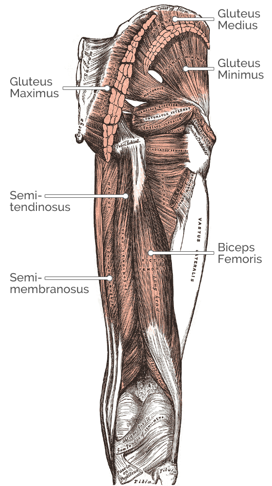
Muscles involved in knee flexion:
Biceps Femoris
Origin: Tuberosity of the ischium, femoral linea
aspera
Insertion: Head of the fibula articulating with
the back of the lateral tibial condyle
Other actions: Knee flexion, laterally rotates
knee when knee is flexed
Semimembranosus
Origin: Ischial tuberosity
Insertion: Medial condyle of tibia
Other actions: Knee flexion
Semitendinosus
Origin: Lower Quadrangular part of ischial
tuberosity
Insertion: Pes anserinus - distal tendon of the
semitendinosus, gracilis and sartorius
Other actions: Knee flexion
Knee Flexion
Nerve Anatomy
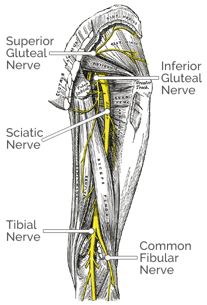
Nerves involved in knee flexion:
Biceps Femoris
Nerve innervation: Tibial nerve (long head)
common peroneal nerve (short head)
Nerve root: L5, S1
Semimembranosus
Nerve: Tibial part of the sciatic nerve
Nerve root: L5, S1, S2
Semitendinosus
Nerve innervation: Tibial part of the sciatic nerve
Nerve root: L5, S1, S2
